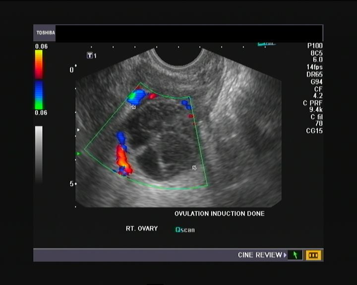
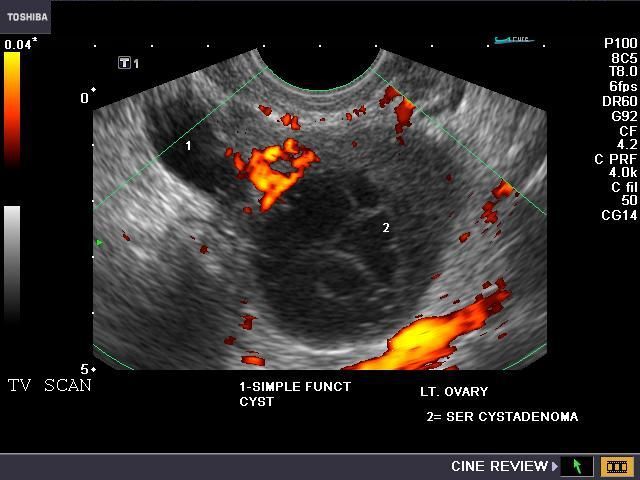
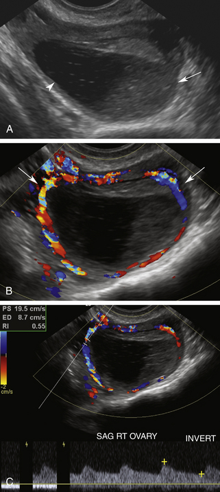
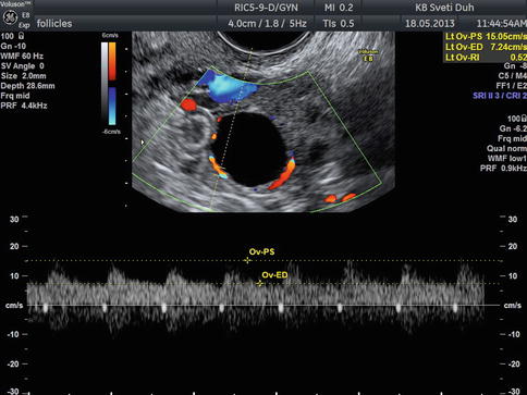
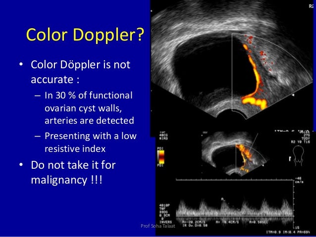
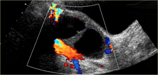
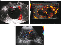
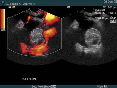
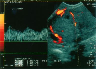
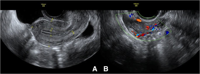
Using the power of 3d ultrasounds doppler evaluation better illuminates the presence of rich vascularity color score 3 4 which is predominant in metastatic cancers as well as the central localization of vessels in a tumor which are important parameters that contribute to the differentiation between benign and malignant ovarian masses.
Early stage ovarian cancer color normal ovary doppler ultrasound.
The early detection of ovarian carcinoma continues to be a formidable challenge and an elusive task.
30 1 ovarian follicles typically achieve a size of 2 to 3 cm before ovulation.
One ovary with stage ia and another ovary in a case with stage ib ovarian cancer were missed.
The cancer is in both ovaries.
Stage 1a the cancer is limited or localized to one ovary.
These data show the ability of transvaginal color doppler sonography to detect ovarian cancer as early as stage i even in asymptomatic women as well as in the morphologically normal ovary.
A small sample is sent to a laboratory where a pathologist examines it under a microscope.
Normal and inconsequential findings.
There are also cancer cells on the.
For ovarian cancer that usually means surgical removal of the mass and one or both ovaries.


